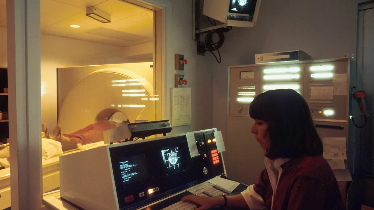
Positron Emission Tomography, or PET, is a cutting-edge medical imaging technique used to observe how tissues and organs function in the body. Unlike traditional imaging methods like X-rays or CT scans that show the structure of organs, PET focuses on capturing the metabolic activity and functioning of cells, providing a deeper insight into health conditions.
PET works by using a small amount of radioactive material, called a tracer, which is injected into the body. This tracer emits positrons, which are detected by a PET scanner to create detailed images of areas with high chemical activity. These areas often correspond to disease, such as tumors in cancer, brain disorders, or heart disease.
One of the most important uses of PET is in cancer detection and monitoring. By highlighting abnormal metabolic activity in cells, PET scans can help identify the location and size of tumors, even in their early stages. Additionally, PET is invaluable in brain imaging, helping doctors diagnose neurological conditions like Alzheimer’s disease and epilepsy.
PET scans are also used to evaluate heart function and detect coronary artery disease. The ability to observe blood flow and metabolic processes in the heart allows doctors to pinpoint blockages or areas of damaged tissue.
Overall, PET is an essential tool in modern medicine, offering precise diagnostic capabilities that guide treatment decisions and improve patient outcomes.
Source : hopkinsmedicine.org
